Imaging Infections: Integrated, multiscale Visualization of Infections and Host Response
Marie Sklodowska Curie European Training Network for fighting infectious Diseases
Infectious diseases are caused by organisms, usually microscopic in size, such as bacteria, viruses, fungi, or parasites that are passed from one person to another. They are a leading cause of death worldwide. To help reduce worldwide mortality, the European training network IMAGE-IN aims to educate a new generation of leading experts in advanced imaging and data analysis methods. It provides insights into molecular imaging, multiscale visualisation of infections and host response to help detect infection and its cause, and determine its severity for personalised treatment. The focus will be on combining data from different imaging modalities to better understand the pathogenesis of difficult-to-treat infections.
About
The European training network IMAGE-IN aims to contribute to the reduction of the worldwide mortality rate caused by infectious diseases by educating a new generation of leading experts in advanced imaging and data analysis methods that will help to better understand and fight infections. The network will provide cutting-edge insights into molecular imaging, multiscale visualization of infections and host response that can help to detect infection, determine its severity and characterize the cause of infection in a time efficient manner to enable personalized treatment. IMAGE-IN actively involves the non-academic sector (SMEs and hospitals) in a unique doctoral training to increase the employability of the IMAGE-IN researchers and to adapt their new skills to the needs of businesses and wider society. While molecular imaging is already common in the clinic, the five research projects target on how data from different imaging modalities can be combined for a better understanding of the pathogenesis of difficult-to-treat infections and ultimately, can lead to faster diagnostics. These domains play essential roles in understanding pathogenesis of infections but also expedite bench-to-bedside translation of new biophotonic and spectroscopic insights (e.g. for diagnostics or later for therapy). The five projects' supervisors have proven track records of success in research and training and bring in perspectives from the public and the private sector. Trainee mobility within the network, in combination with dedication, strong affiliations and technology, creates a highly synergistic framework for success. This assures efficient transfer of excellent academic results to the industry and strengthens Europe's human capital base in R&I through a new generation of entrepreneurial and highly-skilled early career researchers.
ESR Projects

Contact: shibarjun.mandal@leibniz-ipht.de
Supervisors: Ute Neugebauer, Luis Bastiao Silva, Verena Hoerr
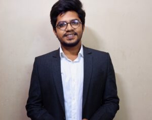
Shibarjun received his bachelor’s in Electrical and Electronic Engineering from the West Bengal University of Technology and a Master’s in Intelligent automation and robotics from Jadavpur University, India in 2015 and 2019 respectively. His master’s degree thesis was on CNN-based anomalies segmentation in endoscopy images. He worked as a project assistant in the Department of Electronics and Telecommunication Engineering, Jadavpur University, on the development of a medical diagnostic device based on olfaction technology. His research interests are medical imaging & processing, AI, ML, and medical devices. Currently, he is doing a Ph.D. degree in the department of Clinical Spectroscopic Diagnostics at Leibniz Institute of Photonic Technology, Jena, Germany.
Shibarjun’s MSCA Project: Localizing the pathogen and monitoring host response in tissue infections
It is often very difficult to distinguish sterile tissue inflammation from tissue infections. My aim is to establish an imaging algorithm that can localize, quantify and characterize the invading pathogen as well as to reveal different levels of host response during the infection process and by this unambiguously detect infections. I will use different imaging modalities will be used and combine their advantages. Exemplarily, I will develop an imaging algorithm for bone infections, in particular haematogenous osteomyelitis. As S. aureus is the major cause of bone infections, I will use a model of haematogenous osteomyelitis to establish imaging modalities and respective data analysis procedures of bone tissue. A translation to human tissue sample is planned.
Mahyasadat Ebrahimi (Leibniz Institute of Photonic Technology Jena / Germany)
Contact: mahyasadat.ebrahimi@leibniz-ipht.de
Supervisors: Rainer Heintzmann, Eduardo Pinho, Verena Hoerr
Mahyasadat received her bachelor’s in Physics from Amirkabir University of Technology (Tehran, Iran) and Master’s in Particle Physics from Semnan University (Semnan, Iran) in 2013 and 2016 respectively. After graduation, she joined as a researcher at TANP company and worked to improve the efficiency of medical devices. Furthermore, she got to focus on image analysis and segmentation. One of the results of this research was the technical note “The Fluo Vision System for Fluorescence Concentration Imaging”. Her research interests are Medical imaging, MRI, and Image processing and segmentation. Currently, she is doing a Ph.D. degree at IPHT and in physical chemistry at the Faculty of Chemistry of the University of Jena, Jena, Germany
Mahyasadat’s MSCA Project: Analysis and visualization of dynamic host responses in animal models with systemic infections by using MRI in-vivo
Severe systemic infection and inflammation, such as sepsis, is described as an inappropriate systemic inflammatory response syndrome. The dysregulated host response results in functional and/or structural tissue damage whose extent is directly correlated to disease severity. There is increasing evidence, that metabolic reprogramming and maladaptation is responsible for organ dysfunction and disease. Also, autoregulation of organ blood flow is reported to be disturbed in critical illness and to vary with cardiac output. However, knowledge of the association of these adverse developments is scarce. I will develop new magnetic resonance imaging (MRI) techniques and establish them to visualize and assess metabolic and haemodynamic organ function in non-invasive longitudinal studies and correlated to molecular imaging of organ dysfunction.
 Rustam Guliev (Leibniz Institute of Photonic Technology Jena/ Germany)
Contact: rustam.guliev@leibniz-ipht.de
Supervisors: Ute Neugebauer, José Luis Oliveira, Claudia Beleites
Rustam has received his specialist degree in Applied Mathematics and Computer Science from Moscow State University, Russia in 2013. From 2013 till 2015 he worked as a developer of Dataware House of one of the biggest telecom companies in Russia. Then, He worked as a research assistant from 2015 till 2022 in Emanuel Institute of Biochemical Physics of Russian Academy of Sciences, Moscow, Russia. Currently, he is doing a Ph.D. degree in Leibniz-Institute of Photonic Technologies.
Project: Characterization of pathogenesis and treatment of infections with obligate intracellular pathogens
Intracellular infections are often difficult to treat, in particular when the pathogens enter persistent states. I will establish new imaging technologies to visualize bacteria and their direct environment inside intact host cells, reveal details on their metabolic state, distinguish different developmental forms and follow antibiotic treatment in a cell culture model. Exemplarily, the imaging technologies will be established for Chlamydia, an obligate intracellular pathogen that causes severe and often persisten infections (high recurrence rate also after antibiotic treatment). Chlamydiae possess a biphasic life cycle and upon stress can enter a persistent state, which – upon now – is very difficult to characterize with conventional methods. In cell culture models of Chlamydia infection the persistent state will be induced and treated with different antibiotics in various formulations. Pathogenesis and treatment will be followed in a time- and dose-dependent manner using different imaging modalities.

Contact: yubrajgupta416@gmail.com
Supervisors: Eduardo Pinho, Rainer Heintzmann, Carlos Costa
Yubraj obtained his bachelor’s degree in Electronics and Communication Engineering from Nepal Engineering College affiliated with Pokhara University, Nepal in June 2016 and his master’s degree in engineering from Chosun University, South Korea in 2019. He served as a research assistant at Chosun University’s Digital Media Computing Laboratory from August 2017 until April 2020. During this time, he published an article on computational neuroscience (focused on Alzheimer’s disease). His research interests include computer vision, medical image processing, multi-modal imaging systems, and spectroscopic imaging modalities. Currently, he is pursuing a PhD in computer engineering at the University of Aveiro in Aveiro, Portugal. The published article can be found here.
Yubraj’s MSCA Project: 3D visualization and correlation from different spectroscopic imaging modalities to define the pathogen niche
There are different techniques to generate 3D microscopy images from hyper-spectral 2D images and depth profiles. All of them need to be optimized to deal with a high amount of data to create a 3D volume. Also, the 3D visualization varies between modalities and requires dedicated software to render the surface. This can be a barrier when you have a heterogeneous environment with images generated from different modalities and the need to visualize it in different software. This problem is addressed in this project and I will propose a common framework for 3D visualization of different microscopy imaging modalities. A plugin-based architecture will allow adding new microscopy modality imaging and preprocessing methods on-demand. Moreover, I will research novel methods for 3D visualization that combine images obtained from different modalities (acquisition techniques). My aim is to generate 3D restauration of microscopy images with improved rendering quality. I will provide a set of artificial intelligence tools, mostly based in deep learning models, to improve knowledge extraction and interaction with 3D microscopy images and will exploit their application in 3D images generated from microscopy imaging. I will exploit the application of this kind of deep learning architectures in 3D microscopy image segmentation. There is also room to apply machine learning methods for identification of mixed patterns in microscope images. In this connection, I will research classification models to identify these patterns which may allow us to recognize different cell types of highly different morphology and patters present in specific diseases.
Rodrigo Escobar Díaz Guerrero (BMD Software – Building the future of healthcare. Ílhavo, Aveiro / Portugal)
Contact: rodediazg@gmail.com
Supervisors: José Luis Oliveira, Jürgen Popp, Thomas Bocklitz, Lina Carvalho
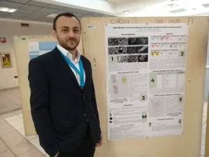
Rodrigo has received a BSc in Computer Engineering and an MSc in Computer Science from the U.A.Q (Autonomous University of Queretaro). For his thesis projects, he worked under the supervision of Dr. Jesús Carlos Pedraza Ortega, performing a Volumetric three-dimensional representation system of moving images, acquired by a depth sensor (2012) and Digitization of solids with fringe projection using phase-shifting profilometry techniques in ARM architectures (2014 -2016). He has given classes at the bachelor level in subjects such as Artificial Intelligence, Operating Systems, Computer Architecture, Automata, and Formal Languages among others in the Technology Institute of Querétaro (2012 -2016). For 4 years, he has been working on developing web pages mainly using the Django framework and several front-end technologies. Furthermore, he had the opportunity to stay 3 months in Durham, England where he developed a protein quantification software for the department of biological sciences at Durham University. During his studies, he won an internal telecontrol contest and was part of the Organizing Committee of the Geekend (event of technological and cultural diffusion).
Rodrigo’s MSCA Project: Collaborative Research Framework for Pathomics
My MSCA project is focused on four main data modalities: (1) Raman spectroscopy: this data modality requires preprocessing tools like, for instance, normalization techniques, spectral axis alignment, background correction/baseline removal, outlier’s removal related with instrumental artifacts, cosmic ray/spike removal or wavelet for denoising purposes, smoothing filters for removal of high frequency components, and extraction of frequency information present in the Raman signal through Fourier Transform methods. One of my most important goals is the separation of relevant signal from the noise part, using intrinsic variable correlations in a given data set. Multivariate analysis is the common approach to archive this. (2) FIB-SEM (Focused Ion Beam Scanning Electron Microscopes): electron microscopy modality for ultrastructural organization of cells and tissues. Datasets generated by this modality can range from gigabytes to hundreds of gigabytes. The framework should provide tool for the extraction of features of interest in order to allow visualization, quantification, and therefore comprehension of their 3D organization. (3) Real-time intravital multimodal imaging: using nonlinear microscopic techniques, such as coherent anti-Stokes-Raman-Scattering (CARS), Two-Photon-Excited Fluorescence (TPEF), Second-Harmonic-Generation (SHG) and Fluorescence lifetime imaging (FLIM). (4) Histopathology: based on whole slide imaging modality. The framework will provide tools for generating, interrogating, and characterizing large volumes of quantitative features from high-resolution tissue images. My project goal is a cloud-based system able to collect pathology imaging studies, extract and indexes features. It will provide tools for extraction of statistical, morphological and spatial metrics/indicators. It will manage the creation of research and educational datasets, and the respective annotation process. Advanced searching mechanism will support multi-source queries and exporting of results (datasets) for feeding artificial intelligence algorithms. The final aim of the project is to develop, evaluate and integrate different image analysis methods in a cloud-based system to allow collaborative pathology imaging research.
The work will be carried out for BMD-Software in close collaboration with the University of Aveiro, where I am enrolled as a doctoral student. At the moment, I am doing a secondment of 10 months at the Leibniz Institute of Photonic Technology in Jena, Germany, for statistical analysis of multimodal data, and plan to stay of 2 months at the University of Coimbra (Prof. Lina Carvalho, FMUC), for my image interpretation.
Local Networks
The research programme of IMAGE-IN is linked to the two profile lines LIGHT and LIFE of the Friedrich-Schiller University of Jena (FSU). The research approach in IMAGE-IN will complement existing programmes and bridge in particular the optical-focussed Abbe School of Photonics and the life-science oriented PhD programmes of the CSCC and the Jena School for Microbial Communication. All these existing graduate programmes support the IMAGE-IN network as IMAGE-IN could fill existing gaps and complement the newly funded German excellence cluster “Balance of the microverse” of the FSU with a strong focus on imaging infections.
Recent Publications
Max Naumann; Natalie Arend; Rustam R. Guliev; Christian Kretzer; Ignacio Rubio; Oliver Werz; Ute Neugebauer:
Label-Free Characterization of Macrophage Polarization Using Raman Spectroscopy
Int. J. Mol. Sci. 2023, Volume 24, Issue 1, 824
https://doi.org/10.3390/ijms24010824
********
Yubraj Gupta; Carlos Costa1, Eduardo Pinho; Luı´s A. Bastião Silva; Rainer Heintzmann: IMAGE-IN: Interactive web-based multidimensional 3D visualizer for multimodal microscopy images
https://doi.org/10.1371/journal.pone.0279825
********
Yubraj Gupta, Carlos Costa, Eduardo Pinho and Luis A. Bastiao Silva:
“Improving the Visualization and Dicomization process for the Stacked Whole Slide Imaging”
https://ieeexplore.ieee.org/document/9669349
********
Rodrigo Escobar Díaz Guerrero, José Luís Oliveira:
Improvements in lymphocytes detection using deep learning with a preprocessing stage”
********
Yubraj Gupta, Carlos Costa, Eduardo Pinho and Luis A. Bastiao Silva:
Dicomization of LSM fluorescence composite microscope image with its bioimaging information
https://www.researchgate.net/publication/352197234_CBMS_2021
********
Christina Ebert; Lorena Tuchscherr; Nancy Unger; Christine Pöllath; Frederike Gladigau; Jürgen Popp; Bettina Löffler; Ute Neugebauer:
“Correlation of crystal violet biofilm test results of Staphylococcus aureus clinical isolates with Raman spectroscopic read‐out”
https://doi.org/10.1002/jrs.6237
********
Yubraj Gupta, Carlos Costa, Eduardo Pinho and Luis A. Bastiao Silva:
A DICOM Standard Pipeline for Microscope Imaging Modalities”
********
Theresa Götz, Marcel Dahms, Johanna Kirchhoff, Claudia Beleites, Uwe Glaser, Jürgen A. Bohnert, Mathias W. Pletz, Jürgen Popp, Peter Schlattmann, Ute Neugebauer:
Automated and rapid identification of multidrug resistant Escherichia coli against the lead drugs of acylureidopenicillins, cephalosporins, and fluoroquinolones using specific Raman marker bands
https://doi.org/10.1002/jbio.202000149
********
Contact
Coordinator
Leibniz Institute of Photonic Technology Jena, Germany
Prof. Ute Neugebauer
BMD Software LTD, Portugal
Prof. Jose Luis Oliveira
Administrative Project Management
Outreach and Communication
Disclaimer
This project has received funding from the European Union’s Horizon 2020 research and innovation programme under the Marie Sklodowska-Curie Grant Agreement 861122.
Find us on EU Community Research and Development Information Service Cordis.


